PKM Monoclonal Antibody
-
货号:CSB-MA018072A0m
-
规格:¥2200
-
图片:
-
 Western Blot
Positive WB detected in: Hela whole cell lysate, MCF-7 whole cell lysate, Jurkat whole cell lysate, NIH/3T3 whole cell lysate
All lanes: PKM antibody at 1:4000
Secondary
Goat polyclonal to Mouse IgG at 1/10000 dilution
Predicted band size: 58 kDa
Observed band size: 58 KDa
Exposure time: 1min -
 Western Blot
Positive WB detected in: Mouse Heart tissue, Mouse Brain tissue, Mouse Skeletal Muscle tissue
All lanes: PKM antibody at 1:4000
Secondary
Goat polyclonal to Mouse IgG at 1/10000 dilution
Predicted band size: 58 kDa
Observed band size: 58 KDa -
 Western Blot
Positive WB detected in: Rat Heart tissue, Rat Spleen tissue, Rat Brain tissue
All lanes: PKM antibody at 1:4000
Secondary
Goat polyclonal to Mouse IgG at 1/10000 dilution
Predicted band size: 55-60 kDa
Observed band size: 55-60 kDa -
 Western Blot
Positive WB detected in: MCF-7 whole cell lysate at 40µg, 20µg, 10µg, 5µg, 2.5µg, 1.25µg, 0.625µg, 0.3125µg
All lanes: PKM antibody at 1:4000
Secondary
Goat polyclonal to Mouse IgG at 1/10000 dilution
Predicted band size: 58 kDa
Observed band size: 58 KDa
Exposure time: 5min -
 Western Blot
Positive WB detected in: MCF-7 whole cell lysate
All lanes: PKM antibody at 1:4000, 1:8000, 1:16000, 1:32000, 1:64000, 1:128000, 1:256000
Secondary
Goat polyclonal to Mouse IgG at 1/10000 dilution
Predicted band size: 58 kDa
Observed band size: 58 KDa
Exposure time: 5min -
 IHC image of CSB-MA018072A0m diluted at 1:400 and staining in paraffin-embedded human tonsil tissue performed on a Leica BondTM system. After dewaxing and hydration, antigen retrieval was mediated by high pressure in a citrate buffer (pH 6.0). Section was blocked with 10% normal goat serum 30min at RT. Then primary antibody (1% BSA) was incubated at 4°C overnight. The primary is detected by a biotinylated secondary antibody and visualized using an HRP conjugated SP system.
 -
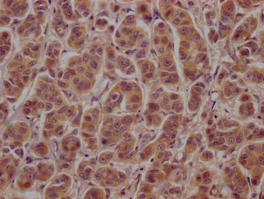
 IHC image of CSB-MA018072A0m diluted at 1:400 and staining in paraffin-embedded human lung cancer tissue performed on a Leica BondTM system. After dewaxing and hydration, antigen retrieval was mediated by high pressure in a citrate buffer (pH 6.0). Section was blocked with 10% normal goat serum 30min at RT. Then primary antibody (1% BSA) was incubated at 4°C overnight. The primary is detected by a biotinylated secondary antibody and visualized using an HRP conjugated SP system.
 -
 IHC image of CSB-MA018072A0m diluted at 1:400 and staining in paraffin-embedded human kidney tissue performed on a Leica BondTM system. After dewaxing and hydration, antigen retrieval was mediated by high pressure in a citrate buffer (pH 6.0). Section was blocked with 10% normal goat serum 30min at RT. Then primary antibody (1% BSA) was incubated at 4°C overnight. The primary is detected by a biotinylated secondary antibody and visualized using an HRP conjugated SP system.
 -
 Immunofluorescence staining of A549 cells with CSB-MA018072A0m at 1:230, counter-stained with DAPI. The cells were blocked in 10% normal Goat Serum and then incubated with the primary antibody overnight at 4°C. The secondary antibody was Alexa Fluor 488-congugated AffiniPure Goat Anti-Mouse IgG(H+L).
 -
 Immunofluorescence staining of Hela cells with CSB-MA018072A0m at 1:230, counter-stained with DAPI. The cells were blocked in 10% normal Goat Serum and then incubated with the primary antibody overnight at 4°C. The secondary antibody was Alexa Fluor 488-congugated AffiniPure Goat Anti-Mouse IgG(H+L).
 -
 Immunofluorescence staining of HepG2 cells with CSB-MA018072A0m at 1:230, counter-stained with DAPI. The cells were blocked in 10% normal Goat Serum and then incubated with the primary antibody overnight at 4°C. The secondary antibody was Alexa Fluor 488-congugated AffiniPure Goat Anti-Mouse IgG(H+L).
 -
 Immunoprecipitating PKM in Hela whole cell lysate
Lane 1: Mouse control IgG instead of CSB-MA018072A0m in Hela whole cell lysate.
Lane 2: CSB-MA018072A0m (1ul) + Hela whole cell lysate (500ug)
Lane 3: Hela whole cell lysate (10ug)
For western blotting, the blot was detected with CSB-MA018072A0m at 1:2000, and a HRP-conjugated Protein G antibody was used as the secondary antibody at 1:2000 -
 Overlay histogram showing Hela cells stained with CSB-MA018072A0m (red line) at 1:100. The cells were incubated in 1x PBS /10% normal goat serum to block non-specific protein-protein interactions followed by primary antibody for 1 h at 4°C. The secondary antibody used was FITC goat anti-mouse IgG(H+L) at 1/200 dilution for 1 h at 4°C. Isotype control antibody (green line) was used under the same conditions. Acquisition of >10,000 events was performed.
 -
 Overlay histogram showing HepG2 cells stained with CSB-MA018072A0m (red line) at 1:100. The cells were incubated in 1x PBS /10% normal goat serum to block non-specific protein-protein interactions followed by primary antibody for 1 h at 4°C. The secondary antibody used was FITC goat anti-mouse IgG(H+L) at 1/200 dilution for 1 h at 4°C. Isotype control antibody (green line) was used under the same conditions. Acquisition of >10,000 events was performed.
-
-
其他:
产品详情
-
产品描述:
CUSABIO has built a mature production system of monoclonal antibodies. Take this PKM monoclonal antibody for example. In the production, the mouses were immunized with Recombinant Human Pyruvate kinase PKM protein at first, and then the spleen cells of the mouse were obtained. Next, used cell hybridization technology to fuse myeloma cells and B lymphocytes in the spleen cells to generate hybridoma. And then screened the obtained hybridoma cells to select the ones that could secrete high-titer antibodies for the production of the PKM monoclonal antibody. This antibody was purified by Protein G and validated in ELISA, WB, IHC, IF, FC, IP.
PKM2 has two isozymes of M type and L type, and M type has M1 and M2 subtypes. M1 is distributed in myocardium, skeletal muscle, and brain tissue; M2 is widely present in tissues during the embryonic period. As the embryo develops, it is gradually replaced by other subtypes in some tissues, and is only highly expressed in cells with a high nucleic acid synthesis rate. , Especially in tumor cells, it is of great significance to the metabolism of cancer and the promotion of tumor formation. At the same time, in the process of tumor progression, due to tumor cell necrosis and metastasis, PKM2 will be released into the blood, and the PKM2 of gastrointestinal tumors It can also be excreted with the stool of cancer patients. Some studies have found that continuous monitoring of PKM2 levels in the plasma of patients with gastric cancer, non-small cell lung cancer, rectal cancer, breast cancer, hepatocellular carcinoma, and renal clear cell carcinoma Comparative analysis shows that the level of PKM2 is significantly increased in tumor patients, which will help the early diagnosis and prognostic judgment of these tumors. Therefore, PKM2 is also used as a very potential new tumor marker. It is expected to become an important anti-tumor drug target. -
产品名称:Mouse anti-Homo sapiens (Human) PKM Monoclonal antibody
-
Uniprot No.:
-
基因名:PKM
-
别名:CTHBP antibody; Cytosolic thyroid hormone-binding protein antibody; KPYM_HUMAN antibody; OIP-3 antibody; Opa-interacting protein 3 antibody; p58 antibody; pkm antibody; PKM1 antibody; PKM2 antibody; Pyruvate kinase 2/3 antibody; Pyruvate kinase muscle isozyme antibody; Pyruvate kinase PKM antibody; THBP1 antibody; Thyroid hormone-binding protein 1 antibody; Tumor M2-PK antibody
-
宿主:Mouse
-
反应种属:Human, Rat, Mouse, Rabbit
-
免疫原:Recombinant Human Pyruvate kinase PKM protein (2-531AA)
-
免疫原种属:Homo sapiens (Human)
-
标记方式:Non-conjugated
-
克隆类型:Monoclonal
-
抗体亚型:IgG1
-
纯化方式:>95%, Protein G purified
-
克隆号:6C3C7
-
浓度:It differs from different batches. Please contact us to confirm it.
-
保存缓冲液:Preservative: 0.03% Proclin 300
Constituents: 50% Glycerol, 0.01M PBS, PH 7.4 -
产品提供形式:Liquid
-
应用范围:ELISA, WB, IHC, IF, FC, IP
-
推荐稀释比:
Application Recommended Dilution WB 1:4000-1:256000 IHC 1:200-1:500 IF 1:150-1:300 FC 1:50-1:200 IP 1µl-4µl -
Protocols:
-
储存条件:Upon receipt, store at -20°C or -80°C. Avoid repeated freeze.
-
货期:Basically, we can dispatch the products out in 1-3 working days after receiving your orders. Delivery time maybe differs from different purchasing way or location, please kindly consult your local distributors for specific delivery time.
产品评价

相关产品
靶点详情
-
功能:Glycolytic enzyme that catalyzes the transfer of a phosphoryl group from phosphoenolpyruvate (PEP) to ADP, generating ATP. The ratio between the highly active tetrameric form and nearly inactive dimeric form determines whether glucose carbons are channeled to biosynthetic processes or used for glycolytic ATP production. The transition between the 2 forms contributes to the control of glycolysis and is important for tumor cell proliferation and survival. In addition to its role in glycolysis, also regulates transcription. Stimulates POU5F1-mediated transcriptional activation. Promotes in a STAT1-dependent manner, the expression of the immune checkpoint protein CD274 in ARNTL/BMAL1-deficient macrophages. Also acts as a translation regulator for a subset of mRNAs, independently of its pyruvate kinase activity: associates with subpools of endoplasmic reticulum-associated ribosomes, binds directly to the mRNAs translated at the endoplasmic reticulum and promotes translation of these endoplasmic reticulum-destined mRNAs. Plays a general role in caspase independent cell death of tumor cells.
-
基因功能参考文献:
- results reveal that nBP1a/PKM2 interaction activates lipid metabolism genes in cancer cells and that Thr-59 phosphorylation of SREBP-1a plays an important role in cancer cell proliferation. PMID: 29514980
- we demonstrated that knockdown of PKM2 can inhibit GC cell proliferation, G1-S phase transition, can, especially, attenuate GC cell migration in vivo and in vitro, per contra, promote the autophagy, which may depend on mediating the PI3K-Akt signaling. PMID: 28588255
- PKM1 activated glucose catabolism and stimulated autophagy/mitophagy, favoring malignancy PMID: 29533781
- miRNA-139-5p inhibited cell proliferation, migration, and glycolysis in GBC, at least in part, by repressing pyruvate kinase M2 PMID: 30105813
- Mammalian target of rapamycin pathway promotes aerobic glycolysis in esophageal squamous cell carcinoma by upregulating pyruvate kinase M2 isoform PMID: 29916308
- PKM2 promotes tumor cell exosome release via phosphorylating protein SNAP23. PMID: 28067230
- proteins such as MMP2 and MMP9 as well as P38 expression were also affected by the PKM2 expression changes. These results proved that PKM2 could be involved in the progression of bladder cancer by mitogen-activated protein kinases signaling pathway. PMID: 30249877
- hypoxic stress in the hepatocellular carcinoma (HCC)cells promoted YAP binding to HIF-1a in the nucleus and sustained HIF-1a protein stability to bind to PKM2 gene and directly activates PKM2 transcription to accelerate glycolysis PMID: 30180863
- overexpressed PKM2 led to increased CCND1 and decreased CDKN1A expression, whereas underexpressed PKM2 led to decreased CCND1 and increased CDKN1A expression in ovarian cancer cells. PMID: 29752805
- Our current study thus unveils a distinct regulatory function of PKM2, providing new options for therapeutic intervention targeting HIV-1-host interactions. PMID: 29607934
- PKM2 as a novel target of RUNX1-ETO and is specifically downregulated in RUNX1-ETO positive AML patients, indicating that PKM2 level might have a diagnostic potential in RUNX1-ETO associated AML. PMID: 28092997
- High M2-PK expression is associated with pancreatic cancer and peri-ampullary cancer. PMID: 29540198
- The activity of PKM2 was indispensable for the development and metastasis of OS. PMID: 29155364
- fFndings demonstrate that mDC activation requires an elevated intrinsic PKM2 level and that PKM2 improves the immune status of patients with SAA by enhancing the functions of mDCs and, consequently, CTLs. PMID: 29636835
- EGFR activation results in c-Src-mediated Cdc25A phosphorylation at Y59, which interacts with nuclear pyruvate kinase M2 (PKM2). PMID: 27485204
- Study demonstrate that PKM2 plays an important role in metabolic activities, as well as in the malignancy of pancreatic ductal adenocarcinoma cells. PMID: 29393401
- Suggest a critical role for pyruvate kinase isozyme M2 in mediating the interaction between pancreatic cancer cells and pancreatic stellate cells. PMID: 29619774
- Results show that PK2 expression level is regulated by HSP90 in hepatocellular carcinoma (HCC). HSP90 enhances PKM2 stability by inducing the phosphorylation of PKM2 at Thr-328. PMID: 29262861
- O-GlcNAcylation is a regulatory mechanism for PKM2 in cancer cells and serves as a bridge between PKM2 and metabolic reprogramming typical of the Warburg effect. PMID: 29229835
- Results indicate that PKM2 is positively correlated with Sp1 expression and demonstrate that Sp1 directly regulates its expression in castration-resistant prostate cancer. PMID: 29094170
- High PKM2 expression is associated with lung adenocarcinoma. PMID: 28489603
- Epigenetic silencing of miR-338 facilitates glioblastoma progression by preventing suppression the PKM2/beta-catenin axis. PMID: 28858851
- this study correlates TuM2PK with tumor size, CRP and CA 15-3 in metastatic breast carcinomas. PMID: 28869444
- PKM2 modulates the glycolysis and extracellular matrix generation, providing the vital role of PKM2 on osteoarthritis (OA) pathogenesis and a novel therapeutic target for OA. PMID: 29356574
- This work suggested that OA increased PKM1/PKM2 ratio, resulting in HNF-4alpha activation and hepatoma differentiation. PMID: 28726775
- the present findings enriched our knowledge by demonstrating a significant association of PKM2 and GLS1 with oxaliplatin-resistance in CRC. PMID: 28498807
- Elevated expression of PKM2 is a prognostic factor for poor gallbladder cancer (GBC) clinical outcomes, implied involving of PKM2 in GBC progression. PMID: 27283076
- PKM2 knockdown resulted in increased p53 expression and prolonged half-life of p53. PKM2 could directly bind with both p53 and MDM2 and promote MDM2-mediated p53 ubiquitination. The dimeric PKM2 significantly suppressed p53 expression compared with the other PKM2 mutants. PMID: 27801666
- Inhibition of the mTOR pathway abolished TGF-beta1-induced EMT and reduced mTOR/p70s6k signaling, which downregulated PKM2 expression. PMID: 28446743
- Study indicates that nitric oxide induces PKM2 nuclear translocation and promotes glycolysis in ovarian cancer cells. PMID: 28380434
- Authors report that AKT directly interacts with PKM2 and phosphorylates it at Ser-202, which is essential for the nuclear translocation of PKM2 protein under stimulation of IGF-1. In the nucleus, PKM2 binds to STAT5A and induces IGF-1-stimulated cyclin D1 expression, suggesting that PKM2 acts as an important factor inducing STAT5A activation under IGF-1 signaling. PMID: 27340866
- The results suggested that targeting PKM2 with an oncolytic adenovirus produced a strong antitumor effect. PMID: 28569774
- our results demonstrated that miR-let-7a inhibits cell proliferation, migration and invasion by down-regulation of PKM2 in cervical cancer PMID: 28415668
- Data show that cytomegalovirus encoded chemokine receptor US28 (US28) signaling in activation of the HIF-1alpha/PKM2 feedforward loop in fibroblasts and glioblastoma cells. PMID: 27602585
- High PKM2 expression is associated urothelial tumorigenesis. PMID: 26992222
- Report that PKM2 is succinylated at lysine 498 and succinylation increases its activity. SIRT5 binds to, desuccinylates and inhibits PKM2 activity. Increased levels of reactive oxygen species (ROS) decreases succinylation and activity of PKM2 by increasing its binding to SIRT5. PMID: 28036303
- over-expression of PKM2 is associated with poor prognosis in most solid cancers and it might be a potentially useful biomarker for predicting cancer prognosis in future clinical applications. PMID: 27911861
- Interdependence of GLO I and PKM2 in the Metabolic shift to escape apoptosis in GLO I-dependent cancer cells PMID: 29225125
- This study demonstrates that lapachol inhibits glycolysis in cancer cells by targeting PKM2. Unlike the previously reported inhibitor shikonin, lapachol does not target mitochondria in tumor cell growth inhibition. PMID: 29394289
- In this study, we indicate that P53 (N340Q/L344R) promotes hepatocarcinogenesis through upregulation of PKM2 PMID: 27167190
- These findings indicate that shRNA-mediated silencing of PKM2 gene promotes apoptosis and inhibits aerobic glycolysis, proliferation, migration, and invasion in colorectal cancer cells. PMID: 28543190
- Due to achieved results concerning expression of PKM2 there is a lack of evidence for its diagnostic and prognostic usage in ovarian cancer. PMID: 29277786
- UCP2 stimulates hnRNPA2/B1, GLUT1 and PKM2 expression and sensitizes pancreatic cancer cells to glycolysis inhibition. PMID: 27989750
- These findings uncover a novel mechanism through which mitochondrial PKM2 phosphorylates Bcl2 and inhibits apoptosis directly. PMID: 28035139
- These findings suggested that PKM2 and GLS might play important roles in the proliferation of hypoxic gastric cancer cells PMID: 29032577
- demonstrated that lincRNA-p21 blunted the prostate cancer cell proliferation and tumorigenic capacity through down-regulation of PKM2 PMID: 28994148
- we demonstrated that SHP-1 dephosphorylates PKM2Y105 to inhibit the Warburg effect and nucleus-dependent cell proliferation, and the dephosphorylation of PKM2Y105 by SHP-1 determines the efficacy of targeted drugs for hepatocellular carcinoma treatment PMID: 26959741
- The PKM2-shRNA group exhibited reduced PKM2 mRNA and protein expression, whereas p53 and p21 expression was increased compared with the blank and empty plasmid groups. PMID: 28746922
- The CARM1-PKM2 axis serves as a metabolic reprogramming mechanism in tumorigenesis. PMID: 29058718
- PKM2 activity is higher in patients with NSCLC than in healthy subjects. The level of PKM2 activity is associated with advanced stage of cancer. PMID: 27683215
显示更多
收起更多
-
亚细胞定位:Cytoplasm. Nucleus.
-
蛋白家族:Pyruvate kinase family
-
组织特异性:Specifically expressed in proliferating cells, such as embryonic stem cells, embryonic carcinoma cells, as well as cancer cells.
-
数据库链接: